Home

generally Transplant soil t1 and t2 values for different tissues disease cross badge

Magnetic Resonance | Radiology Key

PDF] MR imaging: possibility of tissue characterization of brain tumors  using T1 and T2 values. | Semantic Scholar
PDF] MR imaging: possibility of tissue characterization of brain tumors using T1 and T2 values. | Semantic Scholar
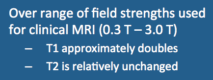
Field strength dependence of T1 & T2 - Questions and Answers ​in MRI

Magnetic Resonance Imaging

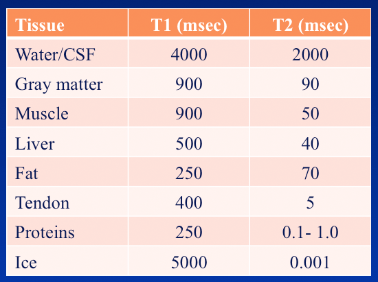
Typical T1 and T2 values for various body tissues* | Download Table

Magnetic Resonance Imaging

1: Typical values of relaxation parameters T1 and T2 (ms) for Hydrogen... |  Download Table
1: Typical values of relaxation parameters T1 and T2 (ms) for Hydrogen... | Download Table

Field strength dependence of T1 & T2 - Questions and Answers ​in MRI

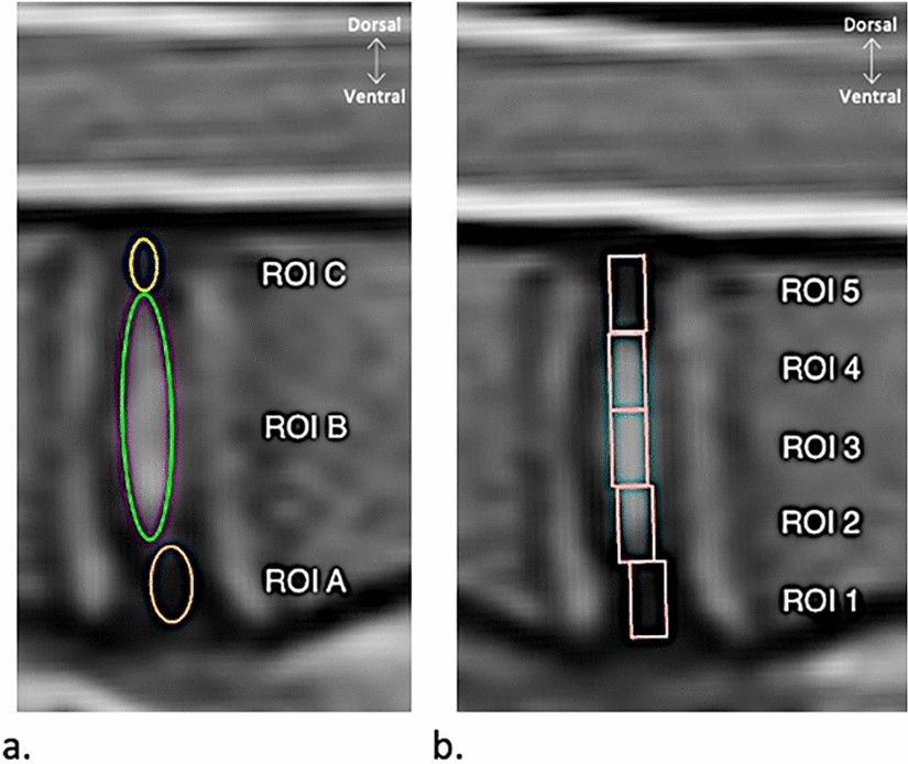
Comparison of MRI T1, T2, and T2* mapping with histology for assessment of  intervertebral disc degeneration in an ovine model | Scientific Reports
Comparison of MRI T1, T2, and T2* mapping with histology for assessment of intervertebral disc degeneration in an ovine model | Scientific Reports

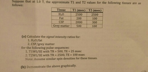
that at 1.0 T, the approximate T1 and T2 values for | Chegg.com

Appearance of various tissues in T1- and T2-weighted images | Download Table

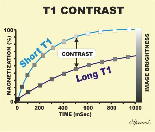
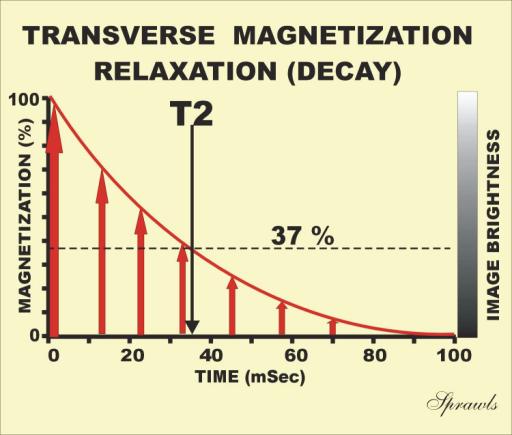
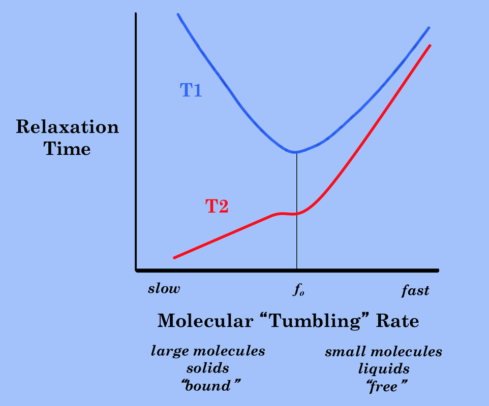
Relaxation time, T1, T2 - Questions and Answers ​in MRI

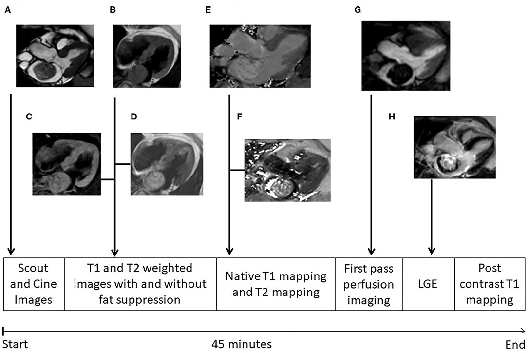
Frontiers | Cardiac Myxomas Show Elevated Native T1, T2 Relaxation Time and  ECV on Parametric CMR
Frontiers | Cardiac Myxomas Show Elevated Native T1, T2 Relaxation Time and ECV on Parametric CMR

Table 1 from Normal tissue quantitative T1 and T2* MRI relaxation time  responses to hypercapnic and hyperoxic gases. | Semantic Scholar
Table 1 from Normal tissue quantitative T1 and T2* MRI relaxation time responses to hypercapnic and hyperoxic gases. | Semantic Scholar

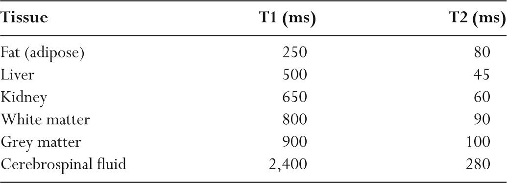
T1 and T2 signal - Radiology Cafe

What are normal relaxation times of tissues at 3 T? | Semantic Scholar

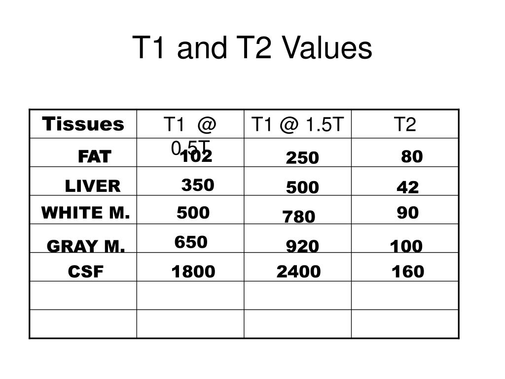
PPT - T1 and T2 Values PowerPoint Presentation, free download - ID:3222942

Relaxation time, T1, T2 - Questions and Answers ​in MRI

Why do body tissues other than the brain have low T2 values for MRI? - Quora

Mapping the Future of Cardiac MR Imaging: Case-based Review of T1 and T2  Mapping Techniques | RadioGraphics
Mapping the Future of Cardiac MR Imaging: Case-based Review of T1 and T2 Mapping Techniques | RadioGraphics

T1 and T2 relaxation times at various anatomical regions at WM and GM... |  Download Table
T1 and T2 relaxation times at various anatomical regions at WM and GM... | Download Table

1. MRI uses differences in T1 and T2 values for the | Download Table

Breast Tissue T1 and T2 Values at 1.5 Tesla | Download Table

Reference T1 and T2 values of the "tissues" in the myocardium phantom |  Download Table
Reference T1 and T2 values of the "tissues" in the myocardium phantom | Download Table

T1 and T2 signal - Radiology Cafe

Suppose that at 1.0 Tesla, the approximate Ti and T2 | Chegg.com